
IDENTIFYING ATTR
Multisystem involvement may be a sign of ATTR
ATTR develops primarily due to the accumulation of amyloid deposits in the heart and other tissues of the body.1-4
- As the disease progresses, multisystem involvement may develop and should be considered a red flag5,6
- Red-flag symptoms can aid in raising clinical suspicion5,6
- A single screening result cannot establish a diagnosis, but may be a warning sign of ATTR
Use the arrows below to explore some of the red-flag symptoms.
Signs that may be identified via echo, EKG, or cMRI:
- Unexplained left ventricular (LV) wall thickening in the absence of hypertension
- Conduction system disease/atrial fibrillation
- Aortic stenosis
- HFpEF in combination with other noncardiac red-flag symptoms
Other clinical symptoms and biomarkers:
- Intolerance/suboptimal response to common cardiovascular medications, including HF treatments*
- Elevated NT-proBNP and troponin
- Shortness of breath
- Edema
*Patients with ATTR-CM can have intolerance to standard medications for heart failure, including ARNi, ACEi, ARB, or β blockers.
ACEi=angiotensin-converting enzyme inhibitor; ARB=angiotensin receptor blocker;
ARNi=angiotensin receptor-neprilysin inhibitor; ATTR-CM=cardiomyopathy of transthyretin-mediated amyloidosis; echo=echocardiography; EKG=electrocardiography; cMRI=cardiac magnetic resonance imaging; HF=heart failure; HFpEF=heart failure with preserved ejection fraction; NT-proBNP=N-terminal prohormone of brain type natriuretic peptide.
- Altered sensation
- Difficulty walking
- Muscle weakness
- Numbness and tingling
- Autonomic nervous system disruptions (eg, GI symptoms, orthostatic hypotension, recurrent UTIs, sexual dysfunction)
GI=gastrointestinal; UTI=urinary tract infection.
- Bilateral carpal tunnel syndrome
- Lumbar spinal stenosis
- Biceps tendon rupture
- Rotator cuff injury
- Trigger finger
- Vitreous opacification
- Glaucoma
- Abnormal conjunctival vessels
- Pupillary abnormalities
- Proteinuria
- Renal failure
Not a comprehensive list of all the symptoms associated with ATTR amyloidosis. Each patient may not experience all of these symptoms or may not experience them at the same time.
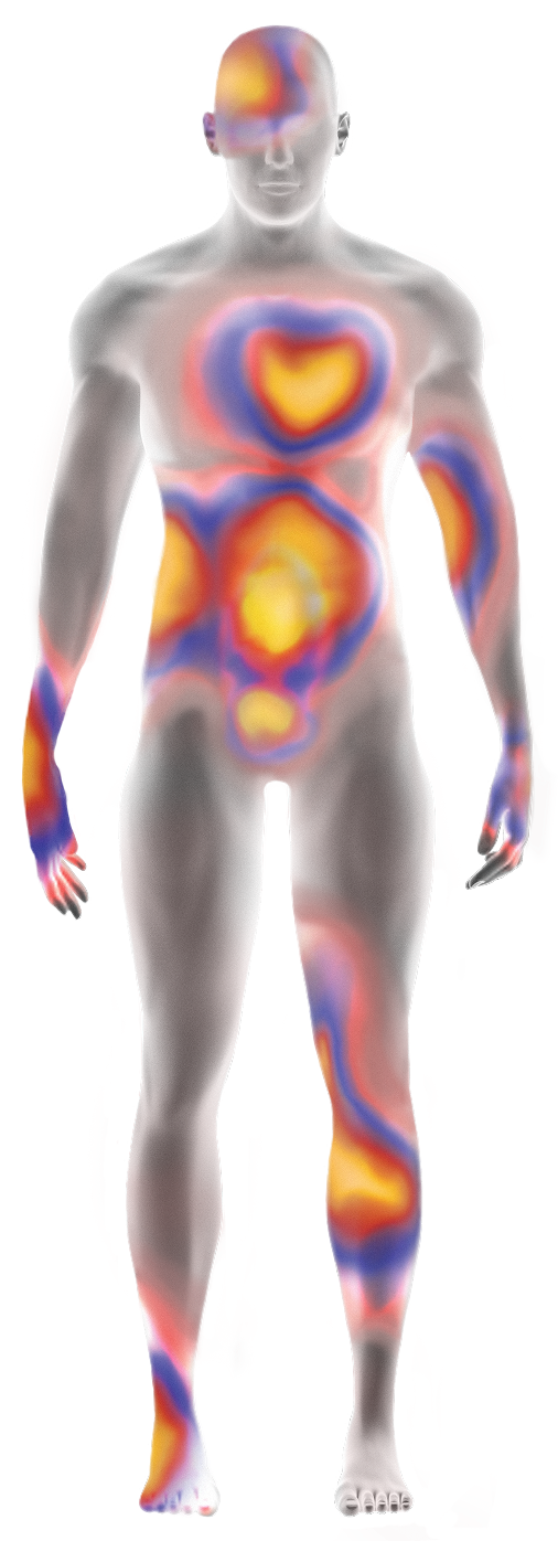
Patients with ATTR experience increasing burden of disease across multiple organ systems as the disease progresses.6,7,15,19
ATTR=transthyretin-mediated amyloidosis.
References:
- Koike H, et al. Biomedicines. 2019;7(1):11.
- Adams D, et al. Neurology. 2015;85(8):675-682.
- Adams D, et al. Curr Opin Neurol. 2016;29(suppl 1):S14-S26.
- Mohty D, et al. Arch Cardiovasc Dis. 2013;106(10):528-540.
- Garcia-Pavia P, et al. Rev Esp Cardiol. 2025;78(4):301-310.
- Kittleson MM, et al. J Am Coll Cardiol. 2023;81(11):1076-1126.
- Kittleson MM, et al. Circulation. 2020;142(1):e7-e22.
- Maurer MS, et al. Circ Heart Fail. 2019;12(9):e006075.
- González-López E, et al. Eur Heart J. 2015;36(38):2585-2594.
- Maloberti A, et al. Int J Cardiol Cardiovasc Risk Prev. 2024;21:200271.
- Dharmarajan K, et al. J Am Geriatr Soc. 2012;60(4):765-774.
- Castaño A, et al. Eur Heart J. 2017;38(38):2879-2887.
- Witteles RM, et al. JACC Heart Fail. 2019 Aug;7(8):709-716.
- Conceição I, et al. J Peripher Nerv Syst. 2016;21(1):5-9.
- Nativi-Nicolau JN, et al. Heart Fail Rev. 2022;27(3):785-793.
- Brito D, et al. Glob Heart. 2023;18(1):1-47.
- Mitter SS, et al. ISA Congress 2020. Poster PT135.
- Maurer MS, et al. J Am Coll Cardiol. 2016;68(2):161-172.
- Rozenbaum MH, et al. Cardiol Ther. 2021;10(1):141-159.