Multiple approved therapeutic options exist for patients with ATTR-CM and hATTR-PN1,2
TTR silencers3,4
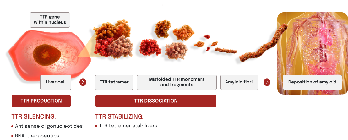
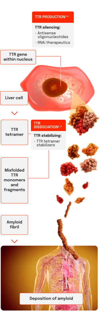
Antisense oligonucleotides (ASOs) are short, chemically modified oligonucleotides that reduce the production of TTR protein
RNA interference (RNAi) therapeutics are double-stranded small interfering RNAs (siRNAs) that reduce the production of TTR protein
TTR stabilizers1,3
TTR tetramer stabilizers are drug molecules that bind to the TTR protein, preventing the dissociation into monomers
Indication(s) vary by specific FDA-approved therapeutic.
Mechanisms to target TTR1-3
TTR silencing works upstream of tetramer formation by suppressing TTR production at its source in the liver. TTR stabilizing works by reducing TTR dissociation and misfolding.
Liver cell image was AI generated.
ATTR=transthyretin-mediated amyloidosis; ATTR-CM=cardiomyopathy of transthyretin-mediated amyloidosis; FDA=Food and Drug Administration; hATTR=hereditary transthyretin-mediated amyloidosis; hATTR-PN=polyneuropathy of transthyretin-mediated amyloidosis; TTR=transthyretin; wtATTR=wild-type transthyretin-mediated amyloidosis.
Links to all outside sites are provided as a reference for our visitors. Alnylam Pharmaceuticals does not endorse and is not responsible for the content on sites that are not owned and operated by Alnylam Pharmaceuticals.
Links to all outside sites are provided as a reference for our visitors. Alnylam Pharmaceuticals does not endorse and is not responsible for the content on sites that are not owned and operated by Alnylam Pharmaceuticals.
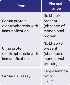
The diagnostic algorithm should always begin with a monoclonal protein screen to assess for AL amyloidosis. If AL amyloidosis is suspected, initiate laboratory testing to assess for monoclonal protein production and evidence of vital organ involvement or dysfunction. In AL amyloidosis, free lambda or free kappa levels are elevated.
ASNC Guidance for 99mTc-PYP Grading14
Two approaches for quantifying cardiac uptake of 99mTc-PYP using planar & SPECT imaging:
Quantitative heart to contralateral lung (H/CL) ratio of uptake at one hour: ATTR-positive if ≥1.5 in the presence of myocardial PYP uptake confirmed on SPECT
Semi-quantitative, visual scoring method to compare myocardial uptake to bone uptake at three hours: ATTR‑positive if ≥Grade 2
Guidance recommends using SPECT or SPECT/CT in addition to planar imaging to confirm diagnosis and avoid false positives.
Biopsy of affected tissues can detect TTR amyloid deposition2,5,10-12
Tissue biopsy can help confirm a diagnosis of ATTR when noninvasive screenings are unavailable or inconclusive.
Standard technique
Biopsy of affected tissue with Congo red staining can detect TTR amyloid deposition.
Use in cardiology
Cardiac tissue biopsy is used when nuclear scintigraphy is negative/equivocal despite a high clinical suspicion of ATTR-CM, or if scintigraphy is unavailable.
Use in neurology
Tissue biopsy is used in the absence of a family history of ATTR, or when genetic testing is inconclusive despite family history, and a high clinical suspicion of hATTR-PN remains.




 LEARN ABOUT A TREATMENT OPTION FOR ATTR‑CM AND hATTR‑PN